Showing posts with label blacksburg. Show all posts

Tuesday, July 13, 2010

Freiberg's Infraction


Freiberg's
Infraction is a condition that affects the lesser metatarsal heads. The most common affected location is the second metatarsal followed by the 3rd 4th then 5th. This condition is also known as AVN (avascular necrosis). Freiberg's Infraction causes a loss of blood supply to the metatarsal head. The condition occurs at the metaphysis of the bone where the nutrient artery of the bone supplies the distal metatarsal. This results in a collapse of the metatarsal head. The condition is more common in females and usually occurs between ages 10-18 and can occur in adulthood.


Classification

Smillie’s classification (1967)


Stage 1: Subtle fracture line through the epiphysis.
Radiographic changes at this stage may be subtle.

Stage 2: Central depression of the articular surface.

Stage 3: Central depression leads to medial and lateral projections at the margins. Plantar hinge remains intact.

Stage 4: Central portion frees from the intact plantar hinge, forming a loose body. Fractures of the medial and lateral projections are present.

Stage 5: Flattening of the metatarsal head with secondary degenerative changes.


Contributing Factors:

Freiberg disease in adolescents is thought to be due to growth disturbances of the epiphysis or apophysis.
  • Vascular insult- an injury of the blood supply to the metatarsal head
  • Traumatic insult- a single acute injury or multiple repetitive injuries

Symptoms
  • Local pain and Tenderness that usually increases with activity
  • Stiffness
  • Limping
Differential Diagnoses
  • Metatarsalgia
  • Morton neuroma
  • Stress fracture
Treatment

Non Surgical:
  • Stiff Soled shoe or Post Op Shoe
  • Non weight-bearing cast
  • Short leg walking cast or CAM boot
  • Rest /Activity modification
  • Steroid injection
Shoe Modifications:
  • Metatarsal pads
  • Rigid shanks
  • Rocker bottom
Surgical Treatment:
  • Simple debridement
Osteotomies:
  • Dorsal closing wedge osteotomies- reorients the plantar intact cartilage to articulate with the proximal phalanx.
  • Shortening osteotomies- metatarsal overloading of the is reduced

Arthroplaty
  • Total joint arthroplasty- Utilizing an implant

Wednesday, June 30, 2010

Tarsal Tunnel Syndrome

Tarsal tunnel syndrome is a condition that is caused by compression of the tibial nerve or its branches.

Tarsal tunnel syndrome is analogous to carpal tunnel syndrome of the wrist.

The tarsal tunnel is a narrow space that lies on the inside of the ankle.

The tunnel is covered with a thick ligament called the flexor retinaculum.

Structures within the tarsal tunnel include:

-Arteries

-Veins
-Tendons
-Nerves


Tarsal tunnel syndrome is a compression on the posterior tibial nerve.

This syndrome produces symptoms anywhere along the path of the nerve.

Possible symptoms include:

-Tingling
-Burning
-Numbness
-Shooting pain
-Paresthesias

Contribute Factors to Tarsal Tunnel:

-Soft tissue masses:
Lipomas-is a benign tumor composed of fatty tissue
-Tendon Sheath Ganglia:
Neoplasms- is an abnormal mass of tissue as a result abnormal proliferation of cells
-Nerve tumors
-Varicose Veins

Work up:

Tinel sign- Percussion of a nerve with radiation of pain along the course of the nerve.

The doctor may order any of the following tests to help with the diagnosis:

-Electromyography (EMG) - A technique for evaluating and recording the electrical activity produced by skeletal muscles
-Nerve conduction velocity (NCV)
-Magnetic resonance imaging (MRI)- In cases of suspected soft-tissue masses and other space-occupying lesions
-Ultrasonography
-Radiography

Conservative Treatment:

-Rest
-Ice
-Non steroidal anti-inflammatory drugs-to help reduce pain and inflammation
-Immobilization- Restricting movement of the foot by wearing a cast or cam boot
-Physical therapy
-Injection therapy
-Orthotic devices. To control any abnormal bio mechanics of the foot


-Surgical Therapy

Tarsal tunnel release surgical intervention may be needed if conservative therapy fails to alleviate pain and symptoms. This procedure is used to decrease pressure on the posterior tibial nerve. Any space occupying lesions may also be excised.

Wednesday, June 9, 2010

Tailor's Bunion


Tailor's bunion is as an acquired lesion that causes chronic pain and swelling over the outer aspect of the distal foot. It is also known as a bunionette and is characterized by a painful prominence on the outer aspect of the foot in the area of the fifth metatarsal head.

These types of lesions were often seen on tailors with a cross-legged sitting posture. The posture resulted in pressure being placed on the lateral side of the foot.

Causes

Extrinsic causes - Commonly chronic
  • Tailors' posture
  • Footwear

Intrinsic causes

Structural Abnormalities
  • Lateral bowing of the metatarsal shaft
  • Enlargement of the metatarsal head
  • Congenital splayfoot

Presentation
  • Symptoms - Painful keratoses on the outer, bottom, or top aspect of the metatarsal head.

Imaging Studies
  • Weight-bearing x rays of both feet
  • Vascular studies are important with patients who have questionable circulation

Treatment

Conservative
  • Padding
  • Shoe modification
  • Orthotic devices
  • Anti-inflammatory medications
  • Corticosteroid injections

Surgical Treatment

  • Can be treated with simple removal of osseous bump when there is no evidence of anglular deformity
  • With an angular deformity or a deviation in the bone is present an osteotomy (cut in the bone) is made and held together with a screw. The level of bone cut varies with the level of the deformity.

Possible Complications

  • Malunion- Incomplete union or union in a faulty position
  • Nonunion- A nonunion occurs when a broken bone does not heal
  • Nerve Injury
  • Joint pain and stiffness
  • Symptomatic hardware
  • Infection
  • Recurrence

Thursday, May 27, 2010

Psoriasis


Psoriasis is a common, chronic, relapsing, inflammatory skin disorder. Psoriasis has a genetic basis and can affect the skin the nails and also joints.

Psoriasis has a tendency to wax and wane.

Flares can be related to systemic, environmental factors, or infection.


Psoriasis most commonly manifests itself on the skin of the elbows, knees, scalp, and lumbosacral areas.


Most psoriatic nail disease occurs in people with clinically evident psoriasis.


Psoriatic arthritis is a chronic inflammatory arthritis that is commonly associated with psoriasis.


Frequency

Approximately 2-3% of people are affected by psoriasis

At least 5% of patients with psoriasis develop psoriatic arthritis

Sex

Psoriasis is slightly more common in women than in men


Symptoms of Psoriasis

The surface of psoriatic lesions often has a layer of dead skin cells that appear as silver scales.

Erythematous(Red) scaly area which can have a sudden onset.

Scaling plaques that itch.

Lesions may be vesicular.

Joint Pain.


Joint Findings

Sausage Digits

Enthesopathy- An inflammation of the insertion points of tendon into bone.

Tendonitis


Nail Findings

Oil drop - a translucent, yellow-red discoloration in the nail bed resembling a drop of oil beneath the nail plate.


Pitting- Pitting is a result of the loss of cells from the surface of the nail plate.


Beau lines- These lines are transverse lines in the nails


Leukonychia- Leukonychia is areas of white nail plate.


Onycholysis Onycholysis is a separation of the nail plate from its attachment to the nail bed.


Causes


Psoriatic lesions are caused by an increase in the skin cells turnover rate.

Genetic factors- Approximately 40% of patients with psoriasis or psoriatic arthritis have first-degree relatives who are affected.

Psoriatic nail disease may be due to a combination of genetic, environmental, and immune factors.


Differentials

  • Seborrheic dermatitis
  • Onychomycosis
  • Squamous cell carcinoma
  • Nummular eczema
  • Lichen planus
  • Lichen simplex chronicus
  • Pustular eruptions


Arthritic Differentials

  • Gout
  • Osteoarthritis
  • Rheumatoid arthritis
  • Septic arthritis


Work Up


Radiographs of affected joints can be help differentiate the type of arthritis.

Psoriatic arthritis- The diagnosis is based primarily on clinical and radiographic findings.

Nail biopsy- A nail biopsy may be obtained to confirm the diagnosis of nail psoriasis


Laboratory studies

Rheumatoid factor (RF)- negative

Erythrocyte sedimentation rate (ESR)- usually normal

Uric acid level- May be elevated in psoriasis


Treatment

Skin Lesions

Topical corticosteroids- Used to reduce plaque formation.


Coal tar- Coal tar is an inexpensive treatment that is available over the counter in shampoos or lotions for use in widespread areas of involvement..

Keratolytic agents- Used to remove scale, to smooth the skin, and to treat hyperkeratosis


Psoriatic arthritis

Nonsteroidal anti-inflammatory drugs- (NSAIDs)

Disease-modifying antirheumatic drugs (DMARDs)

Anti–tumor necrosis factor (TNF)-alpha medications.

Nails
Avulsion therapy -can be used as an alternative therapy for psoriatic nail disease.

Wednesday, May 19, 2010

Gout

Gout is a common disorder of uric acid metabolism. It is a medical condition that usually presents with recurrent attacks of acute inflammatory arthritis. Gout is caused by cellular reaction to uric acid and can lead to deposits of monosodium urate crystals in soft tissues and joints.


Types

Primary gout - May occur alone. Accounts for about 90% of cases of the disease.

Secondary gout- May be associated with other medical conditions or medications. Accounts for about 10% of cases of the disease.


Frequency
Approximately 1% of the general population have gout.

Sex
Predominance- 90% male


Symptoms

Gout is associated with considerable pain.

Acute episodes of gout may incapacitate a patient.

Involved joints typically have the following symptoms: swelling, warmth, erythema, and tenderness.

The first metatarsal phalangeal joint is most commonly affected, however other joints can be involved such as the ankle or the knee.


A tophus deposit may develop in the ear.


Causes

Conditions that may cause acute changes in the level of uric acid and may precipitate a gout attack:

  • Hyperuricemia
  • End-stage renal disease
  • Alcohol ingestion
  • Disorders that cause high cell turnover with release of purines
  • Over consumption of foods high in purines
  • Underexcretion of uric acid - renal insufficiency


Long Term Effects

Untreated chronic tophaceous gout can lead to severe joint destruction.

Deposition of uric acid crystal in the kidneys may produce renal failure or obstruction.


Differential Diagnosis

Cellulitis- A severe inflammation of dermal and subcutaneous layers of the skin.

Gonococcal Arthritis- Is caused by infection with Neisseria gonorrhoeae.

Calcium Pyrophosphate Deposition Disease- Is a type of arthritis caused by the deposition of calcium pyrophosphate crystals.

Rheumatoid Arthritis- Is a chronic systemic inflammatory disease that affects the peripheral joints.

Psoriatic Arthritis- Is a chronic inflammatory arthritis that is commonly associated with psoriasis.


Laboratory Studies

Synovial fluid- The physician may aspirate the involved joint to rule out an infectious arthritis and to confirm a diagnosis of gout.

Serum uric acid.

Uric acid in 24-hour urine sample.


Imaging

Routine radiographs reveal punched-out erosions or lytic areas with overhanging edges. These finding are not acute.


Treatment

Acute gout

  • Indomethacin- is the traditional Nonsteroidal anti-inflammatory drug (NSAID) of choice for acute gout.
  • Colchicine.
  • Corticosteroids- May be indicated in those patients who do not tolerate NSAID or Colchicine.

Chronic gout

  • Probenecid- For patients who are hypoexcreters of uric acid.
  • Allopurinol- For patients who are over producers of uric acid. Allopurinol reduces the generation of uric acid in the body.
  • Uloric- Prevents uric acid production and lowers elevated serum uric acid levels.


Diet

Patients with gout should avoid beer and hard liquor. These elevate levels of uric acid and may precipitate attacks of gout.

High purine foods should be consumed in moderation:
  • Kidney
  • Liver
  • Meats
  • Shellfish

Monday, May 10, 2010

Tinea Pedis

Is a fungal infection of the skin that causes scaling, flaking, and itch of affected areas. It is also known as Ring Worm or Athlete's foot.

Symptoms
The symptoms of athlete's foot or tinea pedis typically include itching and burning of the feet.

The skin may peel or crack with or without any associated pain.

Commonly the rash is localized to the soles of the feet.

Sometimes the flaking skin may spread to the sides and tops of the feet in a moccasin distribution.


The digital interspases may have some moisture, peeling, redness and flaking as well.


Types of Tinea Pedis


T rubrum is the most common cause for tinea pedis.

Trichophyton mentagrophytes, and Epidermophyton floccosum are other causative organisms.


Vesicular tinea pedis-
Usually caused by T mentagrophytes.

This type is characterized by painful, pruritic vesicles most often on the instep.


Interdigital tinea pedis-
Usually caused by T rubrum seen more in hot/ humid environments

This type is characterized by redness, maceration, fissuring, and scaling between toes. It is also associated with itching


Chronic hyperkeratotic tinea pedis-usually caused by T rubrum.
This type is characterized by chronic redness on the bottom of the foot or sides with scaling.


Risk Factors

  • A hot, humid, tropical environment
  • Prolonged use of footwear
  • Hyperhydrosis- Sweating
  • Certain people may have a genetic predisposition to the infection


Work Up

In suspected tinea pedis a KOH (potassium hydroxide) staining may be ordered by the doctor for fungal detection by obtaining a sample of the flaking skin

Fungal culture- may be performed to confirm the diagnosis of tinea pedis. A culture can be used to identify the fungal species.


Treatment

Tinea pedis can be treated with topical or oral antifungals. Some topical medications are over the counter. Topical agents are generally used for 1-6 weeks


Examples of Topical Medication

Topical Imidazoles

  • Clotrimazole 1% (Lotrimin)
  • Econazole 1% cream (Spectazole)
  • Ketoconazole 1% cream (Nizoral)

Topical Pyridones
  • Ciclopirox 1% cream (Loprox)

Topical Allylamines
  • Naftifine 1% cream (Naftin)
  • Terbinafine (Lamisil)

Oral Antifungals

Considered in patients with extensive chronic hyperkeratotic or inflammatory/vesicular tinea pedis
  • Terbinafine (Lamisil)
  • Itraconazole (Sporanox)

Prevention
  • Keeping your feet clean and dry
  • Avoiding prolonged moist environments
  • Disinfecting old shoes
  • Periodic use of anti fungal foot powder in the shoes

Tuesday, May 4, 2010

Morton's Neuroma


Is a painful benign fibrotic enlargement of one of there common digital nerves. It is caused by a shearing force of the adjacent metatarsal bone. It most commonly affects the third common digital nerve. Morton's Neuroma is found to be more common in females. This may be related to the type of shoe gear often worn by females. It is most common in the 4th - 6th decade.


History

Obtaining an accurate history is important to making the diagnosis of Morton's neuroma.

Common Findings
  • Pain in the forefoot and corresponding toes adjacent to the neuroma
  • Pain is usually described as sharp and burning
  • Pain may radiate proximal
  • Numbness and tingling often is observed in the toes adjacent to the neuroma
  • Intermittent pain
  • Massage of the affected area may give some relief
  • Narrow tight high-heeled shoes aggravate the symptoms
  • Patients may feel as though they are walking on a wrinkle in there sock

Exam
  • Firm squeezing of the metatarsal heads with one hand while applying direct pressure to the dorsal and plantar interspace with the other hand may elicit radiating pain.
  • Mulder Sign - A silent palpable click produced by the lateral squeeze test. The neuroma moves between the metatarsals.
  • Passive and active bending of the toe in an upward direction may aggravate symptoms.
  • Sullivan's Sign - Toes adjacent to the affected interspace splay apart on weight bearing.

Imaging
  • Ultrasonography
  • MRI

Differential Diagnosis
  • Stress fracture of the metatarsal
  • Rheumatoid arthritis
  • Hammertoe
  • Metatarsalgia- plantar tenderness over the metatarsal head
  • Neoplasms
  • Metatarsal head osteonecrosis
  • Freiburg osteochondrosis- characterized by interruption of the blood supply of a bone followed by localized bony necrosis.
  • Ganglion cysts
  • Intermetatarsal bursal fluid collections

Treatment

Treatment strategies for Morton's neuroma range from conservative to surgical management.


Conservative

Bio mechanical
  • Orthotics

Medications
  • Injections- Corticosteroid- Anti inflammatory agent
  • Alcohol sclerosing- Causes a chemical neurolysis of the nerve and used as an alternative to surgery for Morton's neuroma
  • NSAID's- Non Steroidal Anti Inflammatory such as Ibuprofen or Naprosyn
  • Tricyclic Antidepressants- Amitriptyline(Elavil)
  • Anticonvulsants- Neurontin (Gabapentin)
  • Pregabalin (Lyrica)
  • Duloxetine (Cymbalta)

Rehabilitation Program
Physical Therapy
  • Cryotherapy-Cold Therapy- Cold may be applied using an ice bag or a cold pack
  • Ultrasonography- Sound waves that are transferred to a specific body area via a round-headed probe. The sound waves travel deep into tissue, creating gentle heat. The heat helps relieve pain and inflammation
  • Deep tissue massage
  • Stretching exercises
  • Phonophoresis- Has been used in an effort to enhance the absorption of topically applied analgesics and anti-inflammatory agents through the therapeutic application of ultrasound

Surgical Intervention
  • Neurectomy- When conservative measures for Morton's neuroma are unsuccessful surgical excision may be beneficial

Tuesday, April 27, 2010

Diabetic Peripheral Neuropathy

Diabetic Peripheral Neuropathy is the presence of symptoms and/or signs of peripheral nerve dysfunction in people with diabetes after exclusion of other causes.
Neuropathies are characterized by a progressive loss of nerve fibers

Noninvasive Testing

  • Nerve conduction studies and Electromyography- A test commonly used to evaluate the function and the ability of electrical conduction of the motor and sensory nerves.
  • Quantitative sensory testing
  • Autonomic Function Tests
  • Monofilament testing - If the patient does not feel the wire at 4 or more at 10 testing sites- the test is positive for neuropathy


Pathophysiology of Diabetic Peripheral Neuropathy

Result from vascular disease:
  • Endothelial dysfunction
  • Deficiency of myoinositol-altering myelin synthesis
  • Chronic hyperosmolarity-causing edema of nerve trunks
  • Increased sorbitol and fructose


Diabetic Neuropathy can contribute to Structural foot deformities

Hammertoes

Bunions

Metatarsal deformities

Charcot foot

Eventual tissue breakdown

The symptoms of peripheral neuropathy include the following

  • Hyperesthesia-Is a condition that involves an abnormal increase in sensitivity to stimuli of the senses.
  • Paresthesia- Is a sensation of tingling, pricking, or numbness of a person's skin. It is more generally known as the feeling of "pins and needles".
  • Dysesthesia-It is defined as an unpleasant, abnormal sense of touch, and it may or may not be, considered as a kind of pain.
  • Radicular pain-Is pain "radiated" along the dermatome (sensory distribution) of a nerve due to inflammation or other irritation
  • Anhydrosis-Means lack of sweating.


Peripheral Neuropathy Signs

  • Loss of sensation in the foot- Results in repetitive stress
  • Injuries and fractures
  • Loss of vibratory and position sense
  • Loss of deep tendon reflexes
  • Trophic ulceration
  • Foot drop


Treatment


Diet

  • Patients with diabetic neuropathy should develop a realistic diet for lowering blood glucose. This should be guided by a nutritionist or doctor

Medicines widely used to help with painful symptoms

  • Selective serotonin and norepinephrine reuptake inhibitors- example:Duloxetine (Cymbalta)
  • Tricyclic antidepressants- example: Amitriptyline (Elavil)
  • Anticonvulsants- example:Gabapentin (Neurontin)
  • Pregabalin (Lyrica)- FDA approved for neuropathic pain associated with diabetic peripheral neuropathy or postherpetic neuralgia.
  • Capsaicin cream (Capsacin)- A natural chemical derived from plants of Solanaceae. This is a topical medicine and is the active component of chili peppers.
  • Neuremedy (Benfotiamine)- For the nutritional management of peripheral neuropathy.

Tuesday, April 20, 2010

Nail Disorders & Nail Surgery

Nails are excellent indicators of disease and provide diagnostic information.

Patients should look for some of the following nail presentations:
  • Discoloration
  • Brittleness
  • Uplifting nail
  • Pitting nail
  • Splitting
  • Striations
  • Nail thinning
  • Ridging
  • Change in nail consistency
  • Change in nail configuration
  • Nail clubbing

Common Nail Problems:


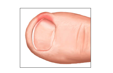
Ingrown nails- Result from an alteration in the proper fit of the nail plate in the usual nail groove. Sharp edges of the lateral nail margin become driven into the skin of the nail groove. The nail essentially becomes a foreign body. An inflammatory response occurs in the involved nail groove. This can lead to redness, swelling, drainage, and development of granulation tissue.


Onychomycosis
- Fungal infections are common, usually caused by dermatophytes.
Common causes: T. rubrum, T. mentogrophytes, and E. Floccosum.
Onychomycosis can be diagnosed by a fungal culture


Anonychia- Is the complete absence of the nail. This condition is a rare congenital anomaly.

Paronychia- Is an infection usually accompanying ingrown nails.

Beau's Lines- Transverse ridges in the nail plate.

Clubbed Digits- marked convexity of nails, with the nail becoming hard, and thick.

Eczematous Conditions- Many types of eczematous dermatitis such as atopic and contact dermatitis can affect the nail folds.

Glomus Tumor
- A purplish tumor that causes pain. The nail bed will appear as a blue-red distortion.

Green Nails- Usually caused by a local Pseudomonas infection

Koilonychia- This describes a spoon shaped nail and describes a characteristic deformity in the form of a concave shape.

Onychauxis-
This is the thickened, elongated, raised irregular nail.
Can be caused by trauma, fungal infection, and nutritional disturbances

Onychogryphosis
- Is an exaggeration of onychauctic condition.

Onychomalacia-
Refers to softness of the nails.

Onycholysis- Detachment of the nail bed from the overlying plate creates a space between nail plate and nail bed.

Splinter Hemorrhages- These are caused by capillary fragility in the longitudinal vessels of the nail bed.


Nail Anatomy


The matrix- is a stratified epithelium that produces hard keratin. The proximal matrix forms the superior nail and the distal matrix forms the lower nail.

Hyponychium- is an epithelial layer of the nail bed

Nail plate- can be separated into zones with predominantly different beginnings. The uppermost layer is generated by the proximal nail fold, the plate by the matrix, and the deepest section of the nail plate is contributed to by the nail folds and bed.

Lunula- is a white semi-lunar area corresponding to the anterior matrix.

Nail bed -consists of the hyponychium and corium over the matrix.


Surgical Nail Procedures:


Nail problems that dictate surgical intervention can include:
Abscess/ Paranychia

Persistent pain

Persistent ingrown nails

Some cases of fungal nails


Procedures:

Phenol and Alcohol Chemical Matrixectomy:

After the toe has been anesthetized, a portion or the whole nail is removed after a sterile preparation of the toe. Next 3 applications of phenol are used at the level of the matrix. The phenol is used to destroy the tissues of the matrix which will prevent further growth of the nail. The area is then flushed with alcohol and a dressing with antibiotic ointment is applied. The patient is given post operative soaking and dressing instructions.

Sodium Hydroxide Matrixectomy

Is a process similar to the phenol and alcohol chemical matrixectomy but uses sodium hydroxide and acetic acid to neutralize. The same criteria apply as for Phenol-Alcohol procedure.

Wednesday, April 7, 2010

Plantar Fasciitis

Plantar fasciitis is one of the most common problems treated in a foot and ankle practice.


Approximately 10% of the United States population experiences bouts of heel pain.


The plantar fascia acts like a windlass mechanism.


The plantar fascia is made up of 3 distinct parts: medial, central, and lateral bands.


It extends from the heel bone to the metatarsal heads.

The plantar fascia is a thick band of tissue in the arch of the foot.


Etiology

Biomechanical dysfunction of the foot is the most common origin of plantar fasciitis.

The pathology is believed to be secondary to the development of microtears in the fascia

There is an inflammation at the fascia at its origin due to repetitive strain of the arch with weight bearing.


Symptoms
Most common complaint is pain in the bottom of the heel.
Patients will typically present with post–static dyskinesia. Pain with the first steps out of bed or periods of rest so it is usually worst in the morning and may improve throughout the day or with more activity.

By the end of the day the pain may be replaced by a dull aching that improves with rest.

Most people complain of increased heel pain after walking for long periods of time.

Generally the most common pain is that elicited upon palpation of the plantar-medial calcaneus

This is at the site of plantar fascial insertion to the heel bone. Pain can occur also at the central and sometimes at the lateral insertion as well.


A tight Achilles tendon can be an adjunctive finding and can contribute to the heel pain. This is known as an Equinus.


Diagnosis
Generally the diagnosis can be made with a good history.

X rays , MRI, and ultrasonography are important modalities to the diagnosis of plantar fasciitis.

X rays may reveal a plantar heel spur, which show the presence of abnormal stresses across the plantar fascia


A heel spur forms in a manner consistent with Wolff’s law. It should be noted that the heel spur is not the cause of the symptoms and therefore does not need specific treatment or removal.

MRI and ultrasonography shows the thickness of the fascia and helps rule out other problems that are not visible with x rays .


Treatment

Nonsurgical treatment include/ Conservative:

  • Rest
  • Icing
  • Stretching
  • Nonsteroidal anti-inflammatory medication such as Ibuprofen
  • Taping/Strapping
  • Orthoses (pre molded or custom-made)
  • Physical Therapy
  • Weight Loss
  • Corticosteroid Injections
  • Night Splints

These treatments should be used in combination.


Walking, running, and jumping sports are associated with plantar fasciitis; restriction of these activities may be necessary.


Surgical:

Severe cases may require surgical intervention if conservative therapy does not improve symptoms.


Extracorporeal shockwave therapy (ESWT) is an alternative treatment for chronic heel pain using acoustic-energy shockwaves


Plantar fascia release—performed by transecting part of the fascia - This is performed through an open incision or performed endoscopically


Another relatively new percutaneous technique is Topaz bipolar radiofrequency microdebridement, which applies a bipolar radiofrequency pulse to the plantar fascia.


Interview with Dr. Feeny

As some of you know, Dr. Feeny recently took her maternity leave from our office. Since she is a Podiatrist I thought it would be valuable to ask her some questions about her feet during her pregnancy and what advice she would give other pregnant women to help take care of their feet.


Hey Dr. Feeny, Thank you so much for letting me be nosey and ask you questions about your feet. First off, did you do anything at the beginning of your pregnancy to prepare for the changes and stress that would be put on your feet in the later months of your pregnancy?

I made sure that I wore supportive shoes throughout my pregnancy. Even if I got up in the middle of the night to go to the bathroom (and believe me that was often) I wore Birkenstock sandals.

At what month of your pregnancy did you notice your feet start hurting?

My feet really did not hurt due to these preventative measures.

That is so great to hear! How did your feet handle having to be on them for a large part of the day?

Well, my feet and ankles would become swollen during the day starting at about month 6. I started wearing compression stockings/support hose which helped.

Did your pregnancy affect your shoe size or the shoes you decided to wear?

I had a hard time tying my shoes that last month so I had to wear slip on shoes. Many women think that their shoes size changes due to weight gain but it is actually due to a hormone. This hormone causes the ligaments to stretch to aid in the childbirth. The ligaments in the feet also stretch which is why it is so important to wear supportive shoes.

Wow! That’s really amazing! I never knew that. So what did you do to ease the pain and symptoms you where experiencing?

I had my hubby rub my feet!! :)

Have you noticed any changes in your feet since you’ve given birth?

The swelling is gone and I can cut my own toenails again.

What advice would you give other pregnant women about taking care of their feet?

Make sure to wear supportive shoes, do not ignore small problems, and do not try to remove any ingrown toenails by yourself.

Thanks again for taking the time to share your experience. It has been really informative!

Tuesday, March 9, 2010

Introducing Dr. Daniel Yeaman


I am pleased to announce that Daniel Yeaman, DPM has joined our team at Shenandoah Podiatry. A native Virginian, Dr. Yeaman recently completed an extensive fellowship in diabetic wounds and limb salvage following a three-year residency, specializing in reconstructive foot and ankle surgery.

His specialties are management and reconstruction of the diabetic foot and ankle. He also treats ingrown nails, skin conditions of the foot such as plantar warts, fungal nails and painful foot conditions such as heel spurs.

He has traveled to Honduras to assist in surgery for children born with foot deformities such as clubfeet and has volunteered locally throughout his education.

In his free time, Dr. Yeaman enjoys hunting, fishing and spending time with his family.

We strive to provide the best care possible for our patients and feel his addition will contribute greatly to our efforts. Please welcome Dr. Yeaman as we all work towards your good health.

Dedicated To Your Healthy Feet,

Dr. Jennifer Feeny

Monday, January 18, 2010

The Strength of Feet: A Remembrance of Martin Luther King Jr.


Today we remember a man who called on the feet of our country to bring about the changes that we needed. We remember those who marched for freedom and stood strong on their beliefs. We look back into history and take into account the miles traveled towards a more equal future for all Americans in our country.

From the beginning to the end of Martin Luther King Jr’s days as one of the most influential people in the civil rights movement, walking was an extremely important part of the campaign to bring about change. From the Montgomery Bus Boycott to the march supporting sanitary waste workers in Memphis, activists used there feet to get their voices heard and heard they were.


One momentous march, the ‘March on Washington for Jobs and Freedom’ has inspired people everywhere to get off the couch and get moving towards the things they believe in. It inspired local marches throughout cities all over the U.S. during the civil rights movements and beyond. Even today we see different groups of people taking to the streets to stand for what they are passionate about and to bring light to inequalities that unfortunately are still present in our world.

Today, take a few steps and remember those who rallied together and used their feet to peacefully make a difference in our world. Take a few steps for those in our history who have fallen fighting for the freedom we enjoy. Today, take a few steps for Martin Luther King Jr. and everything he stood for.


Friday, January 15, 2010

Common Symptoms for Foot Ailments Part 2

Do any of your toenails curl into the fleshy part of your toe causing pain?

You could have an ingrown toenail. This can occur from cutting your toenails rounded at the corners instead of straight across, tight shoes, injuries, toenail fungus, or infections. Do not attempt to cut the toenail out yourself. If you notice your toenail curling even before it is painful call a podiatrist to get this nail removed.

Do you have stinky feet? Do your feet sweat profusely?

You might have Hyperhidrosis or trench foot. This is caused by sweat glands on the bottom of your foot creating more sweat than necessary. What may start off as just an annoyance or unpleasant smell can cause skin to become thick, macerated or painful.

Do you experience a feeling of sharp pain, cramping, or burning in your feet?

You might have a neuroma. A neuroma is when the nerves are pinched by your toe bones. This pinching commonly occurs between the third and forth toes. Tight shoes irritate the symptoms.

Do you have wounds on your feet that do not seem to be healing in a normal amount of time?

You might have chronic wounds or ulcers due to poor circulation or increased pressure on the wound site. These can be very dangerous if they are continuously left untreated. They can become seriously infected or even gangrenous and in severe cases can lead to amputation. Treatment plans can be offered by podiatrists to help the healing process and in most cases can heal in a few months.

Do you have painful growths on the underside of your heel bone? Do the arches of your feet hurt when you get up in the morning?

You might have heel spurs also known as Plantar Fasciitis. This is caused by a strain on the ligaments and muscles attached to your heel bone that lead to the membrane covering the bone to tear. This leaves the ligaments becoming inflamed and the heel bone lacking its natural protective barrier.

If you are experiencing any of these symptoms please call our office and make an appointment today. Don’t live with foot pain any longer! The sooner you get into an office and get your foot pain diagnosed the sooner something can be done about it.

Friday, December 11, 2009

Barefoot Running... The Official Stance

The official stance taken by the American Podiatric Medical Association on barefoot running is as follows:

Barefoot running has become an increasing trend, and a possible alternative or training adjunct to running with shoes. While anecdotal evidence and testimonials proliferate on the Internet and in the media about the possible health benefits of barefoot running, research has not yet adequately shed light on the immediate and long term effects of this practice.

Barefoot running has been touted as improving strength and balance, while promoting a more natural running style. However, risks of barefoot running include a lack of protection--which may lead to injuries such as puncture wounds--and increased stress on the lower extremities. Currently, inconclusive scientific research has been conducted regarding the benefits and/or risks of barefoot running.

The American Podiatric Medical Association, along with the American Academy of Podiatric Sports Medicine, encourages the public to consult a podiatrist with a strong background in sports medicine to make an informed decision on all aspects of their running and training programs.

Barefoot Running

A new trend in running has taken off in recent years though it is not a new concept. People have been kicking off their running shoes to go au nautrale and enjoy the intense connection with the earth that they feel when running this way. Though there has not been a lot of conclusive research done to prove whether or not running shoes are beneficial.

Even with out the concrete evidence showing that barefoot running is better for you, barefoot runners swear by this lifestyle. Barefoot runners believe that with out the obstruction of a running shoe, the brain knows where to place the body and foot in order to run in a way that is most efficient and beneficial to the particular runner. The world was wowed in 1960 when Ethiopian runner Abebe Bikila won his first of many gold medals running barefoot. Of course this was nothing new to him, but it caught everyone else off guard. Barefoot running was also brought to the world’s attention by European runner Bruce Tulloh and American runner’s Charlie Robbinson and Zola Budd.

Research done by Michael Warburton and published in Sportsscience journal entitled ‘Barefoot Running’ which can be found at www.sportsci.org/jour/0103/mw.htm has shown that running shoes increase ankle sprains, Plantar Fasciitis, and other shock related injuries. He also found that less expensive running shoes cause fewer injuries and fewer stressing points on your body than more expensive and advanced running shoes do. According to the article Amby Burfoot wrote in Runnersworld which you can read at http://www.runnersworld.com/article/0,7120,s6-240-319--6728-0,00.html, the brain is much more aware of your body and its surrounds when no shoes are worn.

Despite these findings, running shoes do have their advantages. Most importantly they provide protection. We live in a world filled with dirt, debris, and sharp objects no matter where you are running. Whether that is on a trial, a grassy field, on a side walk, or in a gym, puncture wounds are nothing to take lightly. These wounds can be very dangerous especially if they are not taken care of right away. Running shoes provide support for those with flat feet or high arches. Shoes also protect diabetics from foot injuries. Diabetics are strongly recommended not to try this style of running.

Barefoot running is a very controversial subject. Many people believe that humans should run exactly how they where made and not let anything get in the way of that. Others believe that shoe where invented for a purpose and are extremely important to wear. A third party believes that there is a happy medium between these views and has designed shoes that resemble barefoot running without the risk of puncture wounds. If you are going to start barefoot running make sure to take things very slow and consult your podiatrist before you start. If you don’t have a podiatrist, get one!! And make sure to go to regular check ups. If you are not sure if barefoot running is right for you, do a little research of your own and talk to your podiatrist to decide what is going to be the best running style for you.

Friday, November 27, 2009

The Origin of Shoes


Shoes have come along way since their invention. Some anthropologists believe that the invention of shoes happened as early as 40,000 B.C. These anthropologists noted substantially different toe bone sizes between different generations. The older generations had very big bones; the kind that are associated with a lot of hard work. Then fairly suddenly, the toe bone size got a lot smaller. A very possible explanation to this change is the invention of shoes.


Shoes had an open toed sandal design until about 1600-1200 B.C. when people in colder weather climates created ‘soft shoes’ made out of leather, similar to moccasins, covering the whole foot, and therefore inventing the first close toed shoe. The next major advancement in shoe technology was made by the Egyptians who where the first civilization to make shoes ornate. When the Pharaoh’s where dressed in all their glory, their outfits would be incomplete if they where wearing boring shoes. Shoemaking became a profession in the Egyptian society, making shoes more like jewelry than simply for necessity. Necessity for a hardier shoe brought about the invention of the boot during the ancient Greek era. They needed boots to keep their soldiers protected from different elements in nature and in war. Shoes where not particularly sturdy until about the Middle Ages, when due to the Crusades and the great deal of walking associated with those events, shoes needed to be more durable and more comfortable. Since then shoes have progressed into the styles we know and love today.


In 1850 the first shoe was designed to fit specifically the right or left foot. Soon after that, inventors created special sewing machines that made it able for someone to sew the soles of shoes to the uppers. This new machine expedited the time it took for shoes to be made, leading to the first mass manufacturing of shoes. In 1892 shoes started to look more like the shoes we wear today. Nine rubber companies used their resources and creativity to come up with a new kind of shoe, one that is very quiet and easy to bend. This new rubber sole shoe, a sneaker, was a huge success! Since then, shoes have progresses to fashion as well as necessity, being manufactured and computer engineered instead of handmade, and now mostly made from man made material instead of natural material. Though styles of shoes and techniques of how shoes are made have changed drastically over time, peoples love for shoes and desire to keep inventing haven’t.