Tuesday, April 20, 2010

Nail Disorders & Nail Surgery

Nails are excellent indicators of disease and provide diagnostic information.

Patients should look for some of the following nail presentations:
  • Discoloration
  • Brittleness
  • Uplifting nail
  • Pitting nail
  • Splitting
  • Striations
  • Nail thinning
  • Ridging
  • Change in nail consistency
  • Change in nail configuration
  • Nail clubbing

Common Nail Problems:


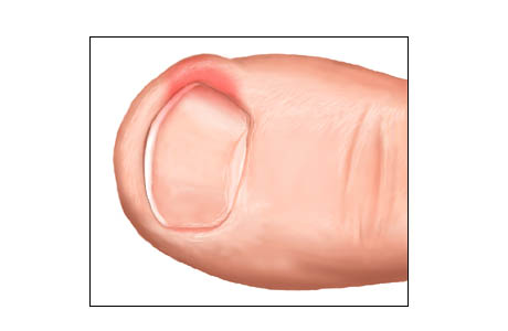
Ingrown nails- Result from an alteration in the proper fit of the nail plate in the usual nail groove. Sharp edges of the lateral nail margin become driven into the skin of the nail groove. The nail essentially becomes a foreign body. An inflammatory response occurs in the involved nail groove. This can lead to redness, swelling, drainage, and development of granulation tissue.


Onychomycosis
- Fungal infections are common, usually caused by dermatophytes.
Common causes: T. rubrum, T. mentogrophytes, and E. Floccosum.
Onychomycosis can be diagnosed by a fungal culture


Anonychia- Is the complete absence of the nail. This condition is a rare congenital anomaly.

Paronychia- Is an infection usually accompanying ingrown nails.

Beau's Lines- Transverse ridges in the nail plate.

Clubbed Digits- marked convexity of nails, with the nail becoming hard, and thick.

Eczematous Conditions- Many types of eczematous dermatitis such as atopic and contact dermatitis can affect the nail folds.

Glomus Tumor
- A purplish tumor that causes pain. The nail bed will appear as a blue-red distortion.

Green Nails- Usually caused by a local Pseudomonas infection

Koilonychia- This describes a spoon shaped nail and describes a characteristic deformity in the form of a concave shape.

Onychauxis-
This is the thickened, elongated, raised irregular nail.
Can be caused by trauma, fungal infection, and nutritional disturbances

Onychogryphosis
- Is an exaggeration of onychauctic condition.

Onychomalacia-
Refers to softness of the nails.

Onycholysis- Detachment of the nail bed from the overlying plate creates a space between nail plate and nail bed.

Splinter Hemorrhages- These are caused by capillary fragility in the longitudinal vessels of the nail bed.


Nail Anatomy


The matrix- is a stratified epithelium that produces hard keratin. The proximal matrix forms the superior nail and the distal matrix forms the lower nail.

Hyponychium- is an epithelial layer of the nail bed

Nail plate- can be separated into zones with predominantly different beginnings. The uppermost layer is generated by the proximal nail fold, the plate by the matrix, and the deepest section of the nail plate is contributed to by the nail folds and bed.

Lunula- is a white semi-lunar area corresponding to the anterior matrix.

Nail bed -consists of the hyponychium and corium over the matrix.


Surgical Nail Procedures:


Nail problems that dictate surgical intervention can include:
Abscess/ Paranychia

Persistent pain

Persistent ingrown nails

Some cases of fungal nails


Procedures:

Phenol and Alcohol Chemical Matrixectomy:

After the toe has been anesthetized, a portion or the whole nail is removed after a sterile preparation of the toe. Next 3 applications of phenol are used at the level of the matrix. The phenol is used to destroy the tissues of the matrix which will prevent further growth of the nail. The area is then flushed with alcohol and a dressing with antibiotic ointment is applied. The patient is given post operative soaking and dressing instructions.

Sodium Hydroxide Matrixectomy

Is a process similar to the phenol and alcohol chemical matrixectomy but uses sodium hydroxide and acetic acid to neutralize. The same criteria apply as for Phenol-Alcohol procedure.

Wednesday, April 7, 2010

Plantar Fasciitis

Plantar fasciitis is one of the most common problems treated in a foot and ankle practice.


Approximately 10% of the United States population experiences bouts of heel pain.


The plantar fascia acts like a windlass mechanism.


The plantar fascia is made up of 3 distinct parts: medial, central, and lateral bands.


It extends from the heel bone to the metatarsal heads.

The plantar fascia is a thick band of tissue in the arch of the foot.


Etiology

Biomechanical dysfunction of the foot is the most common origin of plantar fasciitis.

The pathology is believed to be secondary to the development of microtears in the fascia

There is an inflammation at the fascia at its origin due to repetitive strain of the arch with weight bearing.


Symptoms
Most common complaint is pain in the bottom of the heel.
Patients will typically present with post–static dyskinesia. Pain with the first steps out of bed or periods of rest so it is usually worst in the morning and may improve throughout the day or with more activity.

By the end of the day the pain may be replaced by a dull aching that improves with rest.

Most people complain of increased heel pain after walking for long periods of time.

Generally the most common pain is that elicited upon palpation of the plantar-medial calcaneus

This is at the site of plantar fascial insertion to the heel bone. Pain can occur also at the central and sometimes at the lateral insertion as well.


A tight Achilles tendon can be an adjunctive finding and can contribute to the heel pain. This is known as an Equinus.


Diagnosis
Generally the diagnosis can be made with a good history.

X rays , MRI, and ultrasonography are important modalities to the diagnosis of plantar fasciitis.

X rays may reveal a plantar heel spur, which show the presence of abnormal stresses across the plantar fascia


A heel spur forms in a manner consistent with Wolff’s law. It should be noted that the heel spur is not the cause of the symptoms and therefore does not need specific treatment or removal.

MRI and ultrasonography shows the thickness of the fascia and helps rule out other problems that are not visible with x rays .


Treatment

Nonsurgical treatment include/ Conservative:

  • Rest
  • Icing
  • Stretching
  • Nonsteroidal anti-inflammatory medication such as Ibuprofen
  • Taping/Strapping
  • Orthoses (pre molded or custom-made)
  • Physical Therapy
  • Weight Loss
  • Corticosteroid Injections
  • Night Splints

These treatments should be used in combination.


Walking, running, and jumping sports are associated with plantar fasciitis; restriction of these activities may be necessary.


Surgical:

Severe cases may require surgical intervention if conservative therapy does not improve symptoms.


Extracorporeal shockwave therapy (ESWT) is an alternative treatment for chronic heel pain using acoustic-energy shockwaves


Plantar fascia release—performed by transecting part of the fascia - This is performed through an open incision or performed endoscopically


Another relatively new percutaneous technique is Topaz bipolar radiofrequency microdebridement, which applies a bipolar radiofrequency pulse to the plantar fascia.


Interview with Dr. Feeny

As some of you know, Dr. Feeny recently took her maternity leave from our office. Since she is a Podiatrist I thought it would be valuable to ask her some questions about her feet during her pregnancy and what advice she would give other pregnant women to help take care of their feet.


Hey Dr. Feeny, Thank you so much for letting me be nosey and ask you questions about your feet. First off, did you do anything at the beginning of your pregnancy to prepare for the changes and stress that would be put on your feet in the later months of your pregnancy?

I made sure that I wore supportive shoes throughout my pregnancy. Even if I got up in the middle of the night to go to the bathroom (and believe me that was often) I wore Birkenstock sandals.

At what month of your pregnancy did you notice your feet start hurting?

My feet really did not hurt due to these preventative measures.

That is so great to hear! How did your feet handle having to be on them for a large part of the day?

Well, my feet and ankles would become swollen during the day starting at about month 6. I started wearing compression stockings/support hose which helped.

Did your pregnancy affect your shoe size or the shoes you decided to wear?

I had a hard time tying my shoes that last month so I had to wear slip on shoes. Many women think that their shoes size changes due to weight gain but it is actually due to a hormone. This hormone causes the ligaments to stretch to aid in the childbirth. The ligaments in the feet also stretch which is why it is so important to wear supportive shoes.

Wow! That’s really amazing! I never knew that. So what did you do to ease the pain and symptoms you where experiencing?

I had my hubby rub my feet!! :)

Have you noticed any changes in your feet since you’ve given birth?

The swelling is gone and I can cut my own toenails again.

What advice would you give other pregnant women about taking care of their feet?

Make sure to wear supportive shoes, do not ignore small problems, and do not try to remove any ingrown toenails by yourself.

Thanks again for taking the time to share your experience. It has been really informative!

Tuesday, April 6, 2010

Attention Diabetics

Attention Diabetics, Have you checked your feet recently??

Here is a friendly reminder to check your feet today in case they have been overlooked this past winter. This is an easy exam you can do to see if you need a professional to look at your feet.

Check your feet: for cuts, punctures, irritation, or bruises.
- If you find any of these consult your podiatrist especially if these spots are red, warm or draining.

Check your shoes: make sure you don’t feel any sharp objects in your shoes that would poke your feet.
- If find anything remove it from your shoe and check again.

Check your circulation: push lightly on the tips of your toes that will be pink and reddish in color. The color should change to white when you lift your fingers and then back to pink-red in about 2-3 seconds.
- If the change does not take place right away there might be something affecting your blood flow and you should see your podiatrist asap.

Helpful tip for those with circulation problems… don’t cross your legs when you sit. This will make it even harder for your body to get blood to your feet.

Keep in mind to never put your feet into hot water… only warm water.

Check your feet today!! You never know what you will find.

Wednesday, March 31, 2010

Hammertoes


Digital contraction deformities include hammertoes, clawtoes, and mallet toes.


There are three main causes of digital contracture deformity:

Flexor Stabilization- occurs about >70%. The muscles in the back of the foot and leg fire earlier and longer to stabilize the hypermobile fore foot. This results in overpowering the little muscles in the foot causing the hammertoe. It is possible to see rotation of the 4th and 5th digits with this type of deformity.

Extensor Substitution – is associated with a high arch foot, foot drop, and a weakness of the muscles in the front of the leg.

Flexor Substitution – is the least common cause of digital contracture, and occurs due to weakness of the muscles in the back of the leg.

A mallet toe involves bending of the far toe joint downward and may be associated with a long digit.

Hammertoes involve upward bending of the the first bone in the digit and downward bending of the middle bone in the digit.

The clawtoe involves downward bending of both the middle bone and the far bone of the digit.

Symptoms associated with advanced digital contracture deformity include painful motion, painful hyperkeratotic lesion (Thickened Skin), inability to wear regular shoes, contracted painful toe which is short.

Treatment for claw toes and hammertoes depends on the severity of the deformity.

Goals are to keep the foot comfortable:

If biomechanics is the reason for the hammertoe deformity tan the patient can be fitted for custom-molded orthotics.

Changing to a wider pair of shoes, with more depth in the toe box.

Hammertoe pads or Hammertoe cushions can also be used on hammertoes.

Padding placed under the toes, with a strap that is placed over the toes which helps to straighten the toes.

Pads and cushions can help to alleviate pain and irritation of the toes, these pads however will not change the deformity.

It is possible that the soft tissue structures can begin to tighten. A rigid deformity can develop in such a case.


When conservative care fails and there is considerable pain at the hammertoes, surgery is then considered.

Hammertoe surgery involves straightening the toe through either an arthroplasty by removing a small piece of bone of the digit, or arthrodesis (fusing the joint) using a wire or implant.

Arthroplasty is a minor surgical procedure that may be used to treat hammertoes. In this procedure, the head of the first bone in the digit is removed, allowing the toe to straighten. If the affected toe does not straighten sufficiently after arthroplasty, a number of progressive stepwise soft tissue procedures can be used to attempt to straighten the toe.

In some cases an arthrodesis is necessary. Arthrodesis involves fusing two bones together, typically the first and second bones of the digit. The articular cartilage is removed from each bone. They are then held together with either a pin or an implant.

Following surgery, the patient is placed in a surgical shoe or boot and the patients has limited activity for several weeks.